

If viruses don't exist what about antibodies?
If viruses don't exist what about antibodies?
The baby and the bath water

This post does not describe a technique to show that antibodies exist. It describes a technique that makes use of the phenomena of antibody specificity that is very well characterised. The technique is used millions of times a day in diagnostic and research labs around the world. Antibodies bind to and to localises specific parts of tissues, cells and receptors. If you don’t accept this as enough evidence then I think you would also fail to accept evidence for anything, like the earth being spheroid. In which case there is no point in us debating further.
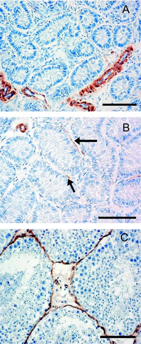
Above image uses an antibody to smooth muscle actin in blood vessel walls and in the myoid cells surrounding the seminiferous tubules in the maturing testes of the harbour porpoise, stained with a brown dye. (1)
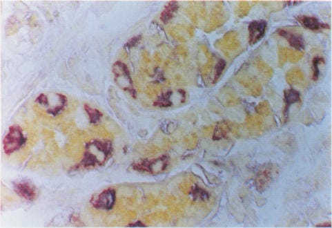
Above imagte shows the secretory canaliculi in the cytoplasm of acid producing parietal cells in the human stomach stained with two dyes, the membrane globulin stained red and the H K ATPase in the proton pump stained blue, co-localising and appearing purple.(2)
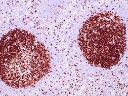
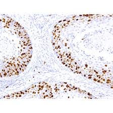
The proliferation marker Ki67 is seen in lymph nodes below.
How is this differentiation and precise staining of receptors, cells and proteins achieved?
NB I do not think there is ever any justification to experiment on other animals, though we may as well learn from what has already been done.
In short mice are injected with a human protein of interest, their serum is screened for reactivity against the protein. In order to make globulins in sufficient concentration to make staining observable, without using billions of mice, they are then sacrificed and B cells from their spleens fused with myeloma cells to make immortal cells lines (hybridomas). Preservatives such as PEG are added. The substance produced from these cell lines is added to a tissue section, and ultimately labelled with a dye. The substance appears to bind and stain only the target protein and leaves all the surrounding proteins untouched.
The localisation of the protein within the morphology of the tissue section provides convincing evidence that the substance is staining the target protein. The staining only occurs where the target protein, is expected to be, in the smooth muscle walls of blood vessels, or in enzyme producing canaliculi for example, and not in any of the other structures or proteins in the tissue section, which provide an internal negative control.
Substances to thousands of different proteins have been produced in this way and the visualisation technique shown to be reproducible literally millions of times a day in diagnostic and research labs around the world.
I don’t see how these staining properties could be achieved using human made chemicals and technology. No amount of fiddling with temperature and pH can make a food colouring or clothes dye accurately locate a part of one cell among a thousand, especially as humans don’t even know the structure and shape of the target proteins they are looking for. Admittedly it is unnatural to inject mammals with proteins and also to form hybridomas; but substances ARE produced by animals that have the remarkable staining properties observed for thousands of target proteins.
I am surprised at the resistance I have met when pointing this out. Chemistry is nowhere near advanced enough to produce this staining. Why should we not think the body makes these specific globulins misnamed ‘antibodies’? Though of course 'measles' antibodies will cross react with 'HIV' proteins etc because there is no such thing as measles or HIV, the proteins the body is producing are the result of an imbalance.
The body has good reason to evolve the ability to ‘recognise’ and bind to all the different antigens that it is exposed to. The body can then judge whether to co-exist with them, such as the bacteria we can’t survive without (they provide skin, gut and brain barriers, are important in digestion and can affect mood), to assimilate them or to expel them.
There also seems to be a tendency to dismiss all microscopy as an artefact. Having worked in pathology for 25 years I know more than anyone what is done to a piece of flesh taken from a living body to make it into a very thin, flat, coloured slice on a microscope slide. However the snapshot of histology is so uniform and consistent that changes between ‘normal’ and ‘abnormal’ are noteworthy. For example, inflammation or a lesion noticed by the patient, seen or felt by the pathologist macroscopically is always (unless someone screwed up) correlated with inflammation, inflammatory cells or lesions microscopically.
Dismissing all of microscopy as artefact is akin to saying that because all historical texts are recorded with bias and are viewed through our own biases, it is not worth bothering to read them at all.
What do ‘antibodies’ do in the body of a sick patient? The paper ‘Measles Virus: A Summary of Experiments Concerned with Isolation, Properties, and Behaviour’ from 1957 (3) is very informative. Enders and Peebles, who sound like butlers in English Manor houses, took samples from people thought to have the measles and added them to kidney cells in culture. They noted 2 cytopathic effects;-

formation of giant cells

and fibroblast-like structures.
The authors claimed that these effects were caused by a virus. However, the absence of cell culture controls using samples from healthy people and from sick patients without the measles, treated in exactly the same way; starving the cells, adding antibiotics and oxidants, makes this claim absurd.
The serum from the sick measles patient was then added to the cell culture. Some globulins in the serum reacted with the combination of proteins in the cell culture and were then referred to as the measles antibodies. The authors were puzzled by these antibodies being present in medium to low titre in 88% of measles history negative patients (asymptomatics anyone?) and decided that in this range nonspecific reactions occurred.
They also postulated that the presence of measles antibodies in normal lab monkeys explained why injecting the ‘virus’, that is cell culture, into their veins and squirting it up their noses did not make them sick.
The authors noted that measles antibodies increased up to 30 fold from the acute phase of the illness to the convalescent phase. This phenomena has been consistently noted for other ‘diseases’ since then. They then added the serum of the convalescing patients to the cell culture and observed that it prevented both types of cytopathic effects seen above. The serum from the acute phase did not. They surmised that neutralising antibodies present in the serum destroyed the virus, and prevented the effects, although if the patient was already convalescing the antibodies were surely too late to be of use!
The disease ‘measles’ is the process of the body detoxifying. In the acute phase, at the beginning of the process, the body creates fever and inflammation, it activates ‘immune’ cells known as mucosal antibodies which may stimulate mucus production in the nose and eyes and develops spots and rashes in order to flush out and expel the toxins through the skin.
The antibodies, produced in the convalescent phase, are binding to the inflammatory cells and substances that were produced in the healing crisis and are bringing the body back down to baseline and balance.
Proteins produced by the stress and oxidation of the cell culture are present in greater amounts in the serum of sick patients than in healthy people and may be these substances that are involved in stimulating the detoxification and healing process. For example the p24 protein associated with ‘HIV’ is present in greater amounts in ‘infected cultures’ than controls and is also seen in pregnancy, inflammation and some cancers. In the case of ‘SARS2’ one of these shock proteins is the so-called spike protein which has been observed in stressed cell cultures since the 90s.
The body does not want to suppress its own detox symptoms in the acute phase, but as the toxins are removed and the patient convalesces, the symptoms need to be reduced and the body returned to a balanced condition. The increase in production of ‘neutralising’ antibodies may bind to these inflammatory proteins, suppress their action and prevent further cytopathic effects, both in cell culture and in the body. The ‘immune system’ and antibodies thus have a role, not in fighting infection, but in healing and homeostasis.
Mike Donio also makes some interesting points about what we call the ‘immune system’ which, if not fighting germs, but may still have a healing and detoxification function:- ‘What is the immune system? Have you really thought about that or just accepted what you have been told in the context that it is your defense against supposed invading pathogenic microbes? Have you considered what else the immune system is doing if there are no contagious diseases?
“Many are talking about this idea of detrimental effects on the immune system resulting from these shots. The claim is usually that there is some sort of immunodeficiency that occurs. What if that’s not entirely incorrect but we are just thinking about it the wrong way? It’s entirely possible that these experimental drugs are causing damage to what we call the ‘immune system.’”
I’m not ready to throw out the ‘immune’ system and the ‘antibody’ baby with germ theory and the ‘viruses’ bath water just yet.
Jo
1.https://pubmed.ncbi.nlm.nih.gov/15379925/ Smooth muscle actin and vimentin as markers of testis development in the harbour porpoise (Phocoena phocoena)
2. https://jcp.bmj.com/content/jclinpath/48/9/832.full.pdf Identification of parietal cells in gastric body mucosa with HMFG-2 monoclonal antibody
3.https://www.ncbi.nlm.nih.gov/pmc/articles/PMC1551024/pdf/amjphnation01086-0004.pdf
4 Nobel laureate lecture 1983 Barbara McClintock
I'm one of those 'throw the antibodies out with germ theory' guys. But good read nevertheless.
Well written!