

Ralph Baric's paper reveals the joke that is peer review;
Ralph Baric's paper reveals the joke that is peer review;
it's worryingly awful


Firstly Airborne transmission of SARS-CoV-2: The world should face the reality
Aside from the fact there is no evidence that disease is transmitted by infectious particles borne by any medium:
they have labelled the large viruses the size of grapefruits; large
and the smaller viruses the size of plums; small
they think that the cartoon needs a key.
Now this sorry excuse for a paper by Ralph Baric SARS-CoV-2 Reverse Genetics Reveals a Variable Infection Gradient in the Respiratory Tract is just ridiculous.
They take wild type (WT) ( a cell culture) and made 2 other viruses (cell cultures with inserts into some RNA fragments of fluorescent dyes, such as GFP, for observation). When the ‘viruses’ (cell cultures) are added to Vero cells they claim to show cytopathic effects (CPE), which denote viral activity and the ability to passage between cells.
However they also showed CPE in the ‘uninfected’, no virus, mock culture. ‘CPE and GFP signals were observed in Vero-E6 cells electroporated with sub-genomic RNA (sgRNA)-N alone (mock)’. We are looking at C.

They also show more ‘viral replication’ in the mock control culture than in the ‘infected’ ones. We are looking at gels of PCR products for the ‘replicated’ M and N ‘genes’ of the 3 ‘viruses’ in B.
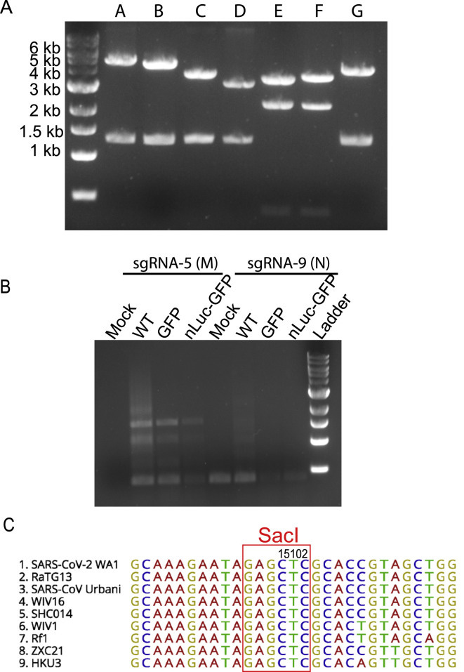
‘B) Amplification SARS-CoV-2 sgRNAs using primers targeting sgRNA-5 (M) and −9 (N). Cellular RNA samples were collected from Vero-E6 cells electroporated with viral RNA transcripts at 20 h. Mock cells were electroporated with SARS-CoV-2 sgRNA-9 (N) alone’…… and had greater expression than the 2 recombinant ‘viruses’ and about the same as WT. They did not test the sgRNA-5 (M) ‘gene’ on the mock so that is why there is no signal.
It shows that the PCR is unsuitable for detecting ‘viruses’ or ‘viral replication’ because it only amplifies short sections of RNA, nothing more.
The insitu hybridisation (ISH) in A and B shows a probe designed for RNA fragments of unknown provenance mistakenly said to be SARS2. This probe is labelled with red dye. Even though the RNA could came from anywhere- the staining in B just looks like a lump of crud to me.
They claim that ‘High-sensitivity RNA in situ mapping revealed the highest angiotensin-converting enzyme 2 (ACE2) expression in the nose with decreasing expression throughout the lower respiratory tract, paralleled by a striking gradient of SARS-CoV-2 infection in proximal (high) versus distal (low) pulmonary epithelial cultures.’
We know that the p132-230 (also called the spike protein) is produced in stressed cell cultures and would be present in what they think of as viruses (which are just cell cultures). It seems to bind to the ACE2 receptor. This would explain their gradient in the respiratory system, if there is one.
The addition of trypsin and proteases (which break down proteins) and the increase in CPE is not surprising, controls weren’t done for this bit. Proteases are also commonly used prior to antibody staining to reveal antigens and enhancing staining- thus making it appear as though there is more ‘viral’ activity- when there isn’t.
The signal from their green florescent protein (GFP) was so weak it required ‘enhancement’ with an antibody. They also observed GFP signal in the mock culture which didn't have a virus with GFP insert added to it!
The paper is a mess of cartoons and images; designed to bamboozle.
What they are basically claiming is that they can show viral passage between cells and viral replication by observing CPE and doing PCR.
However their own paper shows that the mock, with no virus, wild, modified, cloned with GPF insert or otherwise added to it, also showed CPE, GFP signal and had greater expression of alleged viral transcripts amplified by PCR than the alleged viruses.
So they have not shown any virus activity or replication at all.
And the peer review let them get away with saying that they had.
Did they even read it?

Jo
🐒
.
Virology is an embarrassment to the scientific method
Is crud more infective than schmutz or are these just different culture mediums?
Ha Ha